
 Download:


-
Pneumoconiosis, a lung disease primarily characterized by pulmonary fibrosis, occurs due to exposure to dust (1). Phospholipids are pivotal to numerous biological systems, contributing to the formation of cellular lipid bilayers and moderating a host of biological pathways. This moderation is accomplished through the release of signaling molecules such as lysophospholipids, platelet-activating factors, eicosanoids, and diacylglycerides. These molecules participate in the modulation of various processes, including cell proliferation, inflammation, oxidative stress, and neurotransmission, among others (2).
Lipidomics is a potentially useful technique for exploring lipid metabolism and metabolite-related biomarkers in complex respiratory diseases (1). Rindlisbacher et al. discovered lysophosphatidylcholines (Lyso PC), via ultra-high-performance liquid chromatography paired with high-resolution mass spectrometry (UHPLC-HRMS), that could serve as a potential biomarker in the serum of idiopathic pulmonary fibrosis (IPF) patients (3). Using a similar lipidomics analysis, Montesi et al. identified that 22∶4 lysophosphatidic acid (Lyso PA) was significantly elevated in the plasma and exhaled air condensate (EBC) of IPF patients, suggesting its potential as a biomarker for pulmonary fibrosis progression (4). In another study, Yan et al. pinpointed six potential biomarkers capable of distinguishing IPF patients from control subjects following untargeted lipidomics analysis (5). Peng et al., utilizing an analogous untargeted lipidomics technique on the serum of coal worker’ pneumoconiosis (CWP) patients, found differential metabolites in those exposed to coal dust and CWP patients, primarily related to glycerophospholipid metabolism (6). Further research has suggested a close relationship between phospholipid metabolism and the inflammatory process in pulmonary fibrosis (7).
Numerous targeted lipidomic studies suggest that pneumoconiosis could potentially alter the body’s phospholipid metabolism. Dysregulated phospholipids may engage in the pathogenesis of these diseases and could be significant biomarkers. In this study, we employed ultra-high-performance liquid chromatography-tandem mass spectrometry (UPLC-MS/MS) for a targeted lipidomic analysis of the serum phospholipids from pneumoconiosis patients. The aim is to pave the way for new insights into potential lipid biomarkers for pneumoconiosis.
-
Phospholipid standards with a purity greater than 99.0% were obtained from Sigma-Aldrich (St. Louis, MO, USA). MS grade methanol and ammonium formate were sourced from Thermo Fisher (Houston, TX, USA), while analytical grade dichloromethane was procured from Tong Guang Fine Chemicals (Beijing). Butylated hydroxytoluene (BHT) was acquired from Adamas-Beta (Shanghai). Ultrapure water was used throughout the entire experiment. The preparation of the corresponding phospholipid standard, internal standard (IS) solution, and working curve were performed as dictated by the experiment requirements (8).
-
The current study encompassed a total of 46 male pneumoconiosis patients and 46 male workers with exposure to dust but exhibiting no disease symptoms. Notably, 24 pairs of pneumoconiosis patients and dust-exposed workers were meticulously matched based on age and body mass index (BMI) propensity scores for inclusion in the initial pilot study. The rest of the subjects were included in the validation study. Venous blood samples were collected from all participating subjects using non-additive vessels. The serum was then separated through centrifugation at a speed of 1,664 xg for a duration of 10 minutes and subsequently stored at −80 ℃. This research was approved by both the Ethics Committee of the National Institute of Occupational Health and Poison Control of the Chinese Center for Disease Control and Prevention, China [No. 201721], and the Ethics Committee of Integrated Chinese and Western Medicine, Hubei Provincial Hospital, China [No. 2020011]. All participants signed written informed consent and gave permission for their serum samples to be used in this study.
-
A sample of 100 μL of serum was taken and added to a 1.5 mL centrifuge tube, placed into 10 μL mixed IS application solution, and placed into 890 μL CH3OH/CH2CL2 (2∶1, v/v) containing 0.1%BHT. Subsequently, the entire samples were homogenized by vortexing for 5 minutes and laid still in a refrigerator at 4 ℃ for 15 minutes. The sample was centrifuged at 14,010 xg for 20 min at 4 ℃. Finally, the supernatant was quantitatively transferred to a 1.5 mL injection vial for UPLC-MS/MS analysis.
-
An ACQUITY UPLC system (Waters Corp., Milford, MA, USA) equipped with an ACQUITY UPLC BEH shield RP18 column (1.7 μm, 2.1 mm × 100 mm; Waters Corp., USA) was utilized to perform liquid chromatography. The mobile phase was composed of a methanol/water mixture (5∶95, v/v), which included 10 mmol/L of ammonium formate (Solvent A) and methanol (Solvent B). The gradient schedule was set at a flow rate of 0.4 mL/min and followed the following sequence: for the initial minute, it contained 60% of Solvent B; from 1 to 1.5 minutes, it gradually increased from 60% to 100% of Solvent B; from 1.5 to 10 minutes, it maintained 100% of Solvent B; from 10 to 10.1 minutes, it decreased quickly from 100% to 60% of Solvent B; and from 10.1 to 12 minutes, it contained 60% of Solvent B. The designated injection volume was 5 μL. During analysis, all samples were maintained at 4 ℃ and the column temperature was held at 50 ℃.
Mass spectrometry was executed employing the TQ-S Micro system (Waters Corp., Milford, MA, USA), with the electrospray ionization (ESI) source functioning in positive and negative ion modes. Lipids such as phosphatidylserine (PS), lysophosphatidylserine (Lyso PS), phosphatidylethanolamine (PE), Lyso PE, phosphatidylglycerol (PG), phosphatidylinositol (PI) were analyzed under positive ion mode (ESI+), whereas phosphatidylcholine (PC), Lyso PC, lysophosphatidic acid (Lyso PA) were assessed under the negative ion mode (ESI−). Targeted lipidomics were explored utilizing multiple reaction monitoring (MRM) modes. Further instrumental configurations were as follows: ion source temperature was set at 150 ℃; desolvation gas temperature at 350 ℃; desolvation gas flow rate, 1,000 L/h; and a capillary voltage of 4 kV for both positive and negative ions. Detailed parameters for the MRM mass spectrometry analysis are provided in
Supplementary Table S1 .The data collection and analysis of UPLC-MS/MS mass spectrometry was conducted using Masslynx 4.1 (Waters Corp., Milford, MA, USA), supplemented by manual peak integration and calibration checks to assure the qualitative and quantitative precision of each compound analyzed. To enhance data analysis, quantitative data underwent preprocessing — substances with less than an 85% detection rate were omitted and half of the detection limit was utilized to simulate and fill missing values.
-
The Student’s t-test and Rank-sum test were employed to detect substantial differences in phospholipids between the two groups. A combination of principal component analysis (PCA) and orthogonal partial least squares-discriminant analysis (OPLS-DA) was executed, and variable importance of projection (VIP) values were subsequently calculated to isolate differential phospholipids. Phospholipids of significant difference were identified, based on the standard of VIP>1.0 and P<0.05. Additionally, binary logistic classification was utilized to construct a suitable diagnostic model. The receiver operator characteristic curve (ROC) analysis was used to identify potential biomarkers.
The multivariate statistical analysis was conducted using SIMCA software (version 14.1; Umetrics, Scania, Sweden), while the univariate statistical analysis was executed through SPSS software (version 26.0; SPSS Inc, Chicago, IL, USA). A P-value of less than 0.05 was deemed statistically significant. Furthermore, data visualization was facilitated by GraphPad Prism software (version 8.0.2; CA, USA).
-
Table 1 outlines the characteristics of both the pilot and validation study subjects, all of whom were male. In the pilot study, the case and control groups had mean ages of 43.42 and 43.08 years respectively, and mean BMI (kg/m2) of 22.04 and 21.82 respectively. No statistically significant variances in age, BMI, or smoking status were observed between these two groups. Notably, the case group comprised of 18 silicosis and 6 CWP patients. This included 5 instances of stage I pneumoconiosis and 19 of stage III.
Demographic
characteristicsPilot study Validation study Cases (n=24) Controls (n=24) t/χ2 value P-value Cases (n=22) Controls (n=22) t/χ2 value P-value Male (n) 24 24 22 22 Age (years) 43.42±8.42 43.08±8.21 0.139 0.890 53.23±6.13 33.41±3.95 12.756 0.000 Smokers (%) 54.17 54.17 0.000 1.000 63.63 63.63 0.000 1.000 BMI (kg/m2) 22.04±2.42 21.82±2.45 0.325 0.747 21.08±2.28 24.66±2.53 −4.937 0.000 Table 1. Characteristics of the study subjects.
Regarding the validation study subjects, the case and the control groups reported contrasting mean ages, at 53.23 and 33.41 years respectively, and in their mean BMIs (kg/m2) of 21.08 and 24.66 respectively, establishing statistically substantial differences in both age and BMI. However, the smoking status between the two groups showed no significant statistical discrepancy. The case group consisted of 16 silicosis patients, 6 CWP patients, and this included 1 case of stage I pneumoconiosis, 1 case of stage II, and 20 cases of stage III pneumoconiosis.
-
The study performed quantitative analysis of 22 phospholipids present in the serum of 46 pneumoconiosis patients and 46 dust-exposed workers using UPLC-MS/MS methodology. Of these, 16 phospholipids were detected. The initial examination (Figure 1A–E) revealed substantial reductions in serum levels of 16:0 Lyso PC, 18:0–18:1 PG, 18:0–18:1 PE, 18:0 PE, and 18:1 Lyso PE within the case group compared to the control group; these reductions were statistically significant.
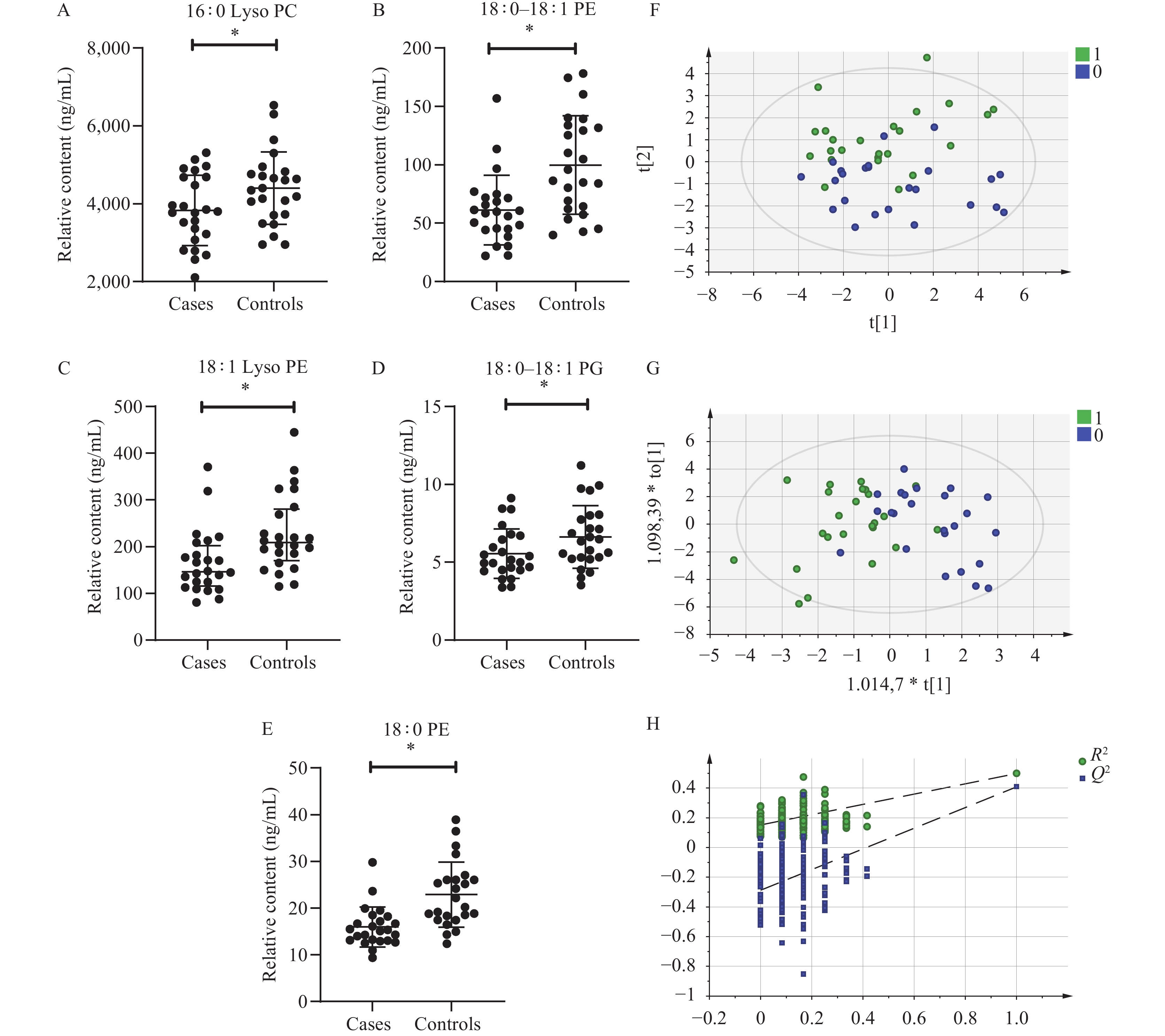 Figure 1.
The univariate and multivariate analysis results between two groups. (A–E) The univariate statistical analysis results. (F) The mode of PCA between the two groups. (G) The mode of OPLS-DA between the two groups. (H) The permutation tests with 200 response sorting for the OPLS-DA model.
Notes: In panels A–E, the single dimensional statistical analysis results: Six differential PLs between the two groups. In panel F, R2X=0.655, Q2=0.197. “1” denotes the case group; “2” denotes the control group. In panel G, R2X=0.506, R2Y=0.499, Q2Y=0.409. “1” denotes the case group; “2” denotes the control group. In panel H, R2=(0.0, 0.164), Q2=(0.0, −0.269).
Abbreviation: Lyso PC=lysophosphatidylcholines; Lyso PE=lysophosphatidylethanolamine; PE=phosphatidylethanolamine; PG=phosphatidylglycerol.
* P<0.05.
-
The PCA highlighted a clear differentiation in serum phospholipids between the two groups, though some overlap was observed (Figure 1F). The OPLS-DA is a supervised pattern recognition approach. This analysis facilitated a distinct separation trend in serum phospholipids between the two groups, further emphasizing substantial differences in serum phospholipid metabolism profiles (Figure 1G). Permutation tests with 200 response permutations confirmed the robustness of the OPLS-DA model (Figure 1H). Notably, the VIP of 18∶0 PE, 18∶0–18∶1 PE, 18∶1 Lyso PE, 18∶0–18∶1 PG, 16∶0 PC, 16∶0–18∶1 PC, 18∶0 PC, and 18∶0–18∶1 PC all exceeded 1.
-
In the preliminary study, using the selection criteria of P<0.05 and VIP>1, four distinct phospholipids were identified: 18∶0 PE, 18∶0–18∶1 PE, 18∶1 Lyso PE, and 18∶0–18∶1 PG. Using pneumoconiosis patients as the dependent variable (where 0=dust-exposed workers, 1=pneumoconiosis patients) and important phospholipids with P<0.05 and VIP>1 (including 16∶0 Lyso PC, 16∶0 PC, 16∶0–18∶1 PC, 18∶0–18∶1 PC, 18∶0 PC, 18∶0 PE, 18∶0–18∶1 PE, 18∶1 Lyso PE, 18∶0 PA, and 18∶0–18∶1 PG) as independent variables, a binary logistic regression analysis was conducted. The resulting diagnostic model equation was Y=1.533+0.010X1−0.304X2, where X1 signifies the concentration of 16∶0 PC and X2 refers to the concentration of 18∶0 PE. Subsequently, an ROC curve analysis of the four distinct phospholipids and the diagnostic model was conducted to investigate their potential efficacy as lipid biomarkers. Notably, the area under the curve (AUC) of 18∶0 PE, 18∶0–18∶1 PE, and 18∶1 Lyso PE exceeded 0.7, indicating a substantial diagnostic value for pneumoconiosis. A diagnostic model with an AUC exceeding 0.8 suggested an enhanced diagnostic performance (Figure 2A, B, E).
 Figure 2.
The ROC curve analysis results for screening and validation of potential lipid biomarkers for pneumoconiosis. (A) ROC curve for significantly different phospholipids in the pilot study. (B) ROC curve for the diagnostic model in the pilot study. (C) ROC curve for 18∶0–18∶1 PE, 18∶0 PE, and 18∶1 Lyso PE in the validation study. (D) ROC curve for the diagnostic model in the validation study. (E) Results of ROC curve analysis.
Abbreviation: Lyso PE=lysophosphatidylethanolamine; PE=phosphatidylethanolamine; PG=phosphatidylglycerol; ROC=receiver operating characteristic; AUC=area under the curve.In the preliminary study, phospholipids and diagnostic models with AUC>0.7 were filtered, and their efficiency as potential lipid biomarkers was duly verified in the validation study. Our findings suggest that 18∶0 PE and 18∶1 Lyso PE exhibited an AUC<0.7, indicating a deficient ability to distinguish pneumoconiosis patients among healthy populations exposed to dust. Conversely, 18∶0–18∶1 PE and diagnostic model, which showed AUC>0.7, demonstrated substantial screening efficiency in the validation studies. These factors, therefore, may serve as potential lipid biomarkers in diagnosing pneumoconiosis (Figure 2C–E).
-
This study utilized a targeted lipidomics analysis to investigate the serum of patients with pneumoconiosis. The findings highlighted significant differences in phospholipid metabolism when compared to healthy workers exposed to dust. Specifically, variances were identified in the expression levels of 18∶0 PE, 18∶0–18∶1 PE, and 18∶1 Lyso PE between the two groups. Furthermore, diagnostic models, dependent upon 16∶0 PC and 18∶0 PE, were developed and exhibited exceptional screening efficiency. The follow-up validation study confirmed that 18∶0–18∶1 PE and the diagnostic model may serve as potential lipid biomarkers for pneumoconiosis.
Lyso PC significantly contributes to the emergence and progression of inflammation (6). Our study has shown that serum levels of 16∶0 Lyso PC were notably lower in pneumoconiosis patients compared to dust-exposed workers. These findings align with the results of a lipid metabolomics study performed on silica dust-exposed rats (9). Conversely, PE has been known to foster an anti-fibrotic phenotype in normal human lung fibroblasts and ameliorate bleomycin-induced pulmonary fibrosis in mice (10). The serum levels of 18∶0–18∶1 PE and 18∶0 PE examined in our research were found to be lower in pneumoconiosis patients in comparison to dust-exposed workers. Such decrease in PE may have implications on the progression of pulmonary fibrosis (11). Lyso PE, on the other hand, may be associated with anti-inflammatory outcomes (12). Our study indicated lower serum levels of 18∶1 Lyso PE in pneumoconiosis patients compared to dust-exposed workers. PG is recognized for its antibacterial and immunosuppressive effects (13–14). Our findings revealed lower serum levels of 18∶0–18∶1 PG in pneumoconiosis patients compared to dust-exposed workers. Such decrease in PG could potentially amplify inflammation (15). Various phospholipids may exert pro-inflammatory or anti-inflammatory effects on the health status of pneumoconiosis patients via different pathways; however, their precise mechanisms warrant further investigation.
To the best of our knowledge, this is the pioneering study applying targeted lipidomics to analyze phospholipid metabolism in pneumoconiosis patients, thus paving the way for novel biomarker research in pneumoconiosis diagnosis. This study, however, does harbour certain limitations. First, the small sample size reduces the robustness of our findings. Additionally, our validation study was hampered due to an age disparity between the cases and their younger controls, and a lack of an external control group consisting of non-dust-exposed healthy individuals. Expanding the sample size and introducing an external control group could be a promising avenue for subsequent validation studies. Second, the representation of different stages and types of pneumoconiosis in our case group was suboptimal. Future research should thus delve deeper into the alterations in phospholipid metabolism across diverse stages and types of pneumoconiosis, thereby unearthing potential lipid biomarkers that could facilitate early detection, monitor disease progression, and predict disease prognosis. Third, the number of targeted phospholipid species analyzed in this study was relatively low. In subsequent studies, it would be beneficial to expand the range of targeted phospholipid species, further bolstering the search for potential lipid biomarkers pertinent to pneumoconiosis.
-
No conflicts of interest.
HTML
Chemicals and Materials
Research Study Design and Sampling Methodology
Methodologies for the Preparation of Samples
UPLC-MS/MS Analysis
Statistical Evaluation
Subject
Univariate Statistical Analysis in the Pilot Study
Multivariate Statistical Analysis in the Pilot Study
Screening and Validation of Potential Lipid Biomarkers for Ppneumoconiosis
| Citation: |

|