Advanced Search

Review: Extensive Disturbances of Intracellular Components and Dysfunctions of Biological Pathways in the Brain Tissues During Prion Infection — China’s Studies

View author affiliations
  • Abstract

    The study describes some of the major findings of changes in intracellular components and biological pathways in the brain during prion infection and hypothesizes some important physiological and pathological approaches mainly based on our studies. Omics techniques analysis of messenger RNA (mRNA) and proteins were carried out in the study. Meanwhile, Western blot, immunohistochemistry, and immunofluorescence were used for protein analysis in different signaling pathways. Statistical analyses were used to describe the protein differences in signaling pathways of infected and normal samples. This report reviewed and summarized our studies on the aberrant changes in intracellular components and biological functions in the brains of prion disease (PrD). Omics analyses proposed extensive abnormal alterations of brain mRNAs transcriptions, protein expressions, and post-translational modifications. The molecular disturbances for microtubule instability and depolymerization, the dysregulations of different signals related with neuron loss and synaptic plasticity, the abnormalities of mitochondrial and endoplasmic reticulum stress, and disturbance of intracellular reactive oxygen species homeostasis during prion infection were precisely analyzed and reviewed. Aberrant disturbances of numerous biological molecules and signals in brain tissues were found during prion infection.

  • loading...
  • Funding: Supported by National Key R&D Program of China (2020YFE0205700), SKLID Development Grant (2021SKLID503, 2021SKLID504, 2019SKLID401), and National Natural Science Foundation of China (81772197, 81630062, 81401670)
  • [1] Tian C, Liu D, Chen C, Xu Y, Gong HS, Chen C, et al. Global transcriptional profiling of the postmortem brain of a patient with G114V genetic Creutzfeldt-Jakob disease. Int J Mol Med 2013;31(3):676 − 88. http://dx.doi.org/10.3892/ijmm.2013.1239CrossRef
    [2] Tian C, Liu D, Sun QL, Chen C, Xu Y, Wang H, et al. Comparative analysis of gene expression profiles between cortex and thalamus in Chinese fatal familial insomnia patients. Mol Neurobiol 2013;48(1):36 − 48. http://dx.doi.org/10.1007/s12035-013-8426-6CrossRef
    [3] Tian C, Liu D, Xiang W, Kretzschmar HA, Sun QL, Gao C, et al. Analyses of the similarity and difference of global gene expression profiles in cortex regions of three neurodegenerative diseases: sporadic Creutzfeldt-Jakob disease (sCJD), fatal familial insomnia (FFI), and Alzheimer's disease (AD). Mol Neurobiol 2014;50(2):473 − 81. http://dx.doi.org/10.1007/s12035-014-8758-xCrossRef
    [4] Shi Q, Chen LN, Zhang BY, Xiao K, Zhou W, Chen C, et al. Proteomics analyses for the global proteins in the brain tissues of different human prion diseases. Mol Cell Proteomics 2015;14(4):854 − 69. http://dx.doi.org/10.1074/mcp.M114.038018CrossRef
    [5] Shi Q, Chen LN, Lv Y, Zhang BY, Xiao K, Zhou W, et al. Comparative proteomics analyses for 139A and ME7 scrapie infected mice brains in the middle and terminal stages. Proteomics Clin Appl 2017;11(5 − 6):1600113. http://dx.doi.org/10.1002/prca.201600113CrossRef
    [6] Chen LN, Shi Q, Zhang BY, Zhang XM, Wang J, Xiao K, et al. Proteomic analyses for the global S-nitrosylated proteins in the brain tissues of different human prion diseases. Mol Neurobiol 2016;53(8):5079 − 96. http://dx.doi.org/10.1007/s12035-015-9440-7CrossRef
    [7] Chen LN, Sun J, Yang XD, Xiao K, Lv Y, Zhang BY, et al. The brain NO levels and NOS activities ascended in the early and middle stages and descended in the terminal stage in scrapie-infected animal models. Mol Neurobiol 2017;54(3):1786 − 96. http://dx.doi.org/10.1007/s12035-016-9755-zCrossRef
    [8] Wang J, Zhang J, Shi Q, Zhang BY, Chen C, Chen LN, et al. Scrapie infection in experimental rodents and SMB-S15 cells decreased the brain endogenous levels and activities of Sirt1. J Mol Neurosci 2015;55(4):1022 − 30. http://dx.doi.org/10.1007/s12031-014-0459-4CrossRef
    [9] Maimaitiming A, Xiao K, Hu C, Chen J, Yang XH, Zhou DH, et al. Aberrant decrease of the endogenous SIRT3 and increases of acetylated proteins in scrapie-infected cell line SMB-S15 and in the brains of experimental mice. ACS Chem Neurosci 2019;10(10):4293 − 302. http://dx.doi.org/10.1021/acschemneuro.9b00341CrossRef
    [10] Li XL, Dong CF, Shi S, Wang GR, Li Y, Wang X, et al. The octarepeat region of hamster PrP (PrP51-91) enhances the formation of microtubule and antagonize Cu2+-induced microtubule-disrupting activity. Acta Biochim Biophys Sin (Shanghai) 2009;41(11):929 − 37. http://dx.doi.org/10.1093/abbs/gmp088CrossRef
    [11] Dong CF, Shi S, Wang XF, An R, Li P, Chen JM, et al. The N-terminus of PrP is responsible for interacting with tubulin and fCJD related PrP mutants possess stronger inhibitive effect on microtubule assembly in vitro. Arch Biochem Biophys 2008;470(1):83 − 92. http://dx.doi.org/10.1016/j.abb.2007.11.007CrossRef
    [12] Li XL, Wang GR, Jing YY, Pan MM, Dong CF, Zhou RM, et al. Cytosolic PrP induces apoptosis of cell by disrupting microtubule assembly. J Mol Neurosci 2011;43(3):316 − 25. http://dx.doi.org/10.1007/s12031-010-9443-9CrossRef
    [13] Wang ZY, Tian C, Jing YY, Gong HS, Guo Y, Shi Q, et al. Knockdown of prion protein (PrP) by RNA interference weakens the protective activity of wild-type PrP against copper ion and antagonizes the cytotoxicity of fCJD-associated PrP mutants in cultured cells. Int J Mol Med 2011;28(3):413 − 21. http://dx.doi.org/10.3892/ijmm.2011.688CrossRef
    [14] Zhou RM, Jing YY, Guo Y, Gao C, Zhang BY, Chen C, et al. Molecular interaction of TPPP with PrP antagonized the CytoPrP-induced disruption of microtubule structures and cytotoxicity. PLoS One 2011;6(8):e23079. http://dx.doi.org/10.1371/journal.pone.0023079CrossRef
    [15] Han J, Zhang J, Yao HL, Wang XF, Li F, Chen L, et al. Study on interaction between microtubule associated protein tau and prion protein. Sci China C Life Sci 2006;49(5):473 − 9. http://dx.doi.org/10.1007/s11427-006-2019-9CrossRef
    [16] Han J, Wang XF, Yao HL, Gao C, Li F, Zhang BY, et al. Prion protein inhibits tau-mediated microtubule formation. Neurosci Bull 2005;21(6):398 − 403.
    [17] Wang XF, Dong CF, Zhang J, Wan YZ, Li F, Huang YX, et al. Human tau protein forms complex with PrP and some GSS- and fCJD-related PrP mutants possess stronger binding activities with tau in vitro. Mol Cell Biochem 2008;310(1 − 2):49 − 55. http://dx.doi.org/10.1007/s11010-007-9664-6CrossRef
    [18] Gong HS, Guo Y, Tian C, Xie WL, Shi Q, Zhang J, et al. Reduction of protein kinase MARK4 in the brains of experimental scrapie rodents and human prion disease correlates with deposits of PrPSc. Int J Mol Med 2012;30(3):569 − 78. http://dx.doi.org/10.3892/ijmm.2012.1025CrossRef
    [19] Guo Y, Gong HS, Zhang J, Xie WL, Tian C, Chen C, et al. Remarkable reduction of MAP2 in the brains of scrapie-infected rodents and human prion disease possibly correlated with the increase of calpain. PLoS One 2012;7(1):e30163. http://dx.doi.org/10.1371/journal.pone.0030163CrossRef
    [20] Chen JM, Gao C, Shi Q, Shan B, Lei YJ, Dong CF, et al. Different expression patterns of CK2 subunits in the brains of experimental animals and patients with transmissible spongiform encephalopathies. Arch Virol 2008;153(6):1013 − 20. http://dx.doi.org/10.1007/s00705-008-0084-zCrossRef
    [21] Chen JM, Gao C, Shi Q, Wang GR, Lei YJ, Shan B, et al. Casein kinase II interacts with prion protein in vitro and forms complex with native prion protein in vivo. Acta Biochim Biophys Sin (Shanghai) 2008;40(12):1039 − 47. http://dx.doi.org/10.1111/j.1745-7270.2008.00486.xCrossRef
    [22] Wang ZY, Shi Q, Wang SB, Tian C, Xu Y, Guo Y, et al. Co-expressions of casein kinase 2 (CK2) subunits restore the down-regulation of tubulin levels and disruption of microtubule structures caused by PrP mutants. J Mol Neurosci 2013;50(1):14 − 22. http://dx.doi.org/10.1007/s12031-012-9845-yCrossRef
    [23] Meng G, Tian C, Wang H, Xu Y, Zhang BY, Shi Q, et al. Remarkable reductions of PAKs in the brain tissues of scrapie-infected rodent possibly linked closely with neuron loss. Med Microbiol Immunol 2014;203(5):291 − 302. http://dx.doi.org/10.1007/s00430-014-0342-5CrossRef
    [24] Sun J, Wang H, Chen LN, Wang J, Lv Y, Yang XD, et al. Remarkable impairment of Wnt/β-catenin signaling in the brains of the mice infected with scrapie agents. J Neurochem 2016;136(4):731 − 40. http://dx.doi.org/10.1111/jnc.13416CrossRef
    [25] Wang H, Tian C, Xu Y, Xie WL, Zhang J, Zhang BY, et al. Abortive cell cycle events in the brains of scrapie-infected hamsters with remarkable decreases of PLK3/Cdc25C and increases of PLK1/cyclin B1. Mol Neurobiol 2013;48(3):655 − 68. http://dx.doi.org/10.1007/s12035-013-8455-1CrossRef
    [26] Wang TT, Tian C, Sun J, Wang H, Zhang BY, Chen C, et al. Down-regulation of brain-derived neurotrophic factor and its signaling components in the brain tissues of scrapie experimental animals. Int J Biochem Cell Biol 2016;79:318 − 26. http://dx.doi.org/10.1016/j.biocel.2016.08.033CrossRef
    [27] Zhang RQ, Chen C, Xiao LJ, Sun J, Ma Y, Yang XD, et al. Aberrant alterations of the expressions and S-nitrosylation of calmodulin and the downstream factors in the brains of the rodents during scrapie infection. Prion 2017;11(5):352 − 67. http://dx.doi.org/10.1080/19336896.2017.1367082CrossRef
    [28] Hu C, Chen C, Chen J, Xiao K, Wang J, Shi Q, et al. The low levels of nerve growth factor and its upstream regulatory kinases in prion infection is reversed by resveratrol. Neurosci Res 2021;162:52 − 62. http://dx.doi.org/10.1016/j.neures.2019.12.019CrossRef
    [29] Yan YE, Zhang J, Wang K, Xu Y, Ren K, Zhang BY, et al. Significant reduction of the GLUT3 level, but not GLUT1 level, was observed in the brain tissues of several scrapie experimental animals and scrapie-infected cell lines. Mol Neurobiol 2014;49(2):991 − 1004. http://dx.doi.org/10.1007/s12035-013-8574-8CrossRef
    [30] Han J, Song QQ, Sun P, Zhang J, Wang X, Song J, et al. Interaction between 14-3-3β and PrP influences the dimerization of 14-3-3 and fibrillization of PrP106-126. Int J Biochem Cell Biol 2014;47:20 − 8. http://dx.doi.org/10.1016/j.biocel.2013.10.013CrossRef
    [31] Shi Q, Song QQ, Sun P, Zhang J, Song J, Chen LN, et al. Infection of prions and treatment of PrP106-126 alter the endogenous status of protein 14-3-3 and trigger the mitochondrial apoptosis possibly via activating Bax pathway. Mol Neurobiol 2014;49(2):840 − 51. http://dx.doi.org/10.1007/s12035-013-8560-1CrossRef
    [32] Xu K, Wang X, Shi Q, Chen C, Tian C, Li XL, et al. Human prion protein mutants with deleted and inserted octarepeats undergo different pathways to trigger cell apoptosis. J Mol Neurosci 2011;43(3):225 − 34. http://dx.doi.org/10.1007/s12031-010-9387-0CrossRef
    [33] Wang X, Dong CF, Shi Q, Shi S, Wang GR, Lei YJ, et al. Cytosolic prion protein induces apoptosis in human neuronal cell SH-SY5Y via mitochondrial disruption pathway. BMB Rep 2009;42(7):444 − 9. http://dx.doi.org/10.5483/BMBRep.2009.42.7.444CrossRef
    [34] Yang XD, Shi Q, Sun J, Lv Y, Ma Y, Chen C, et al. Aberrant alterations of mitochondrial factors drp1 and opa1 in the brains of scrapie experiment rodents. J Mol Neurosci 2017;61(3):368 − 78. http://dx.doi.org/10.1007/s12031-016-0866-9CrossRef
    [35] Gao LP, Xiao K, Wu YZ, Chen DD, Yang XH, Shi Q, et al. Enhanced mitophagy activity in prion-infected cultured cells and prion-infected experimental mice via a pink1/parkin-dependent mitophagy pathway. ACS Chem Neurosci 2020;11(5):814 − 29. http://dx.doi.org/10.1021/acschemneuro.0c00039CrossRef
    [36] Wang X, Shi Q, Xu K, Gao C, Chen C, Li XL, et al. Familial CJD associated PrP mutants within transmembrane region induced Ctm-PrP retention in ER and triggered apoptosis by ER stress in SH-SY5Y cells. PLoS One 2011;6(1):e14602. http://dx.doi.org/10.1371/journal.pone.0014602CrossRef
    [37] Wang SB, Shi Q, Xu Y, Xie WL, Zhang J, Tian C, et al. Protein disulfide isomerase regulates endoplasmic reticulum stress and the apoptotic process during prion infection and PrP mutant-induced cytotoxicity. PLoS One 2012;7(6):e38221. http://dx.doi.org/10.1371/journal.pone.0038221CrossRef
    [38] An R, Dong CF, Lei YJ, Han L, Li P, Chen JM, et al. PrP mutants with different numbers of octarepeat sequences are more susceptible to the oxidative stress. Sci China C Life Sci 2008;51(7):630 − 9. http://dx.doi.org/10.1007/s11427-008-0062-4CrossRef
    [39] Jing YY, Li XL, Shi Q, Wang ZY, Guo Y, Pan MM, et al. A novel PrP partner HS-1 associated protein X-1 (HAX-1) protected the cultured cells against the challenge of H2O2. J Mol Neurosci 2011;45(2):216 − 28. http://dx.doi.org/10.1007/s12031-011-9498-2CrossRef
  • FIGURE 1.  Schema of prion-induced microtubule instability and depolymerization in brains.

    Note: PrPSc and PrP mutants accumulate in neurons and induce the abnormal changes in tubulin, calpain, and many MAPs, leading to microtubule instability and depolymerization.

    Abbreviation: MAPs=microtubule associated proteins; PrPSc=scrapie-like prion protein; PrP=prion protein, TPPP=Tubulin polymerization promoting protein.

    FIGURE 2.  Schema of prion-induced mitochondrial stress, ER stress, and oxidative stress in brains.

    Note: Accumulations of PrPSc and PrP mutants cause aberrant alterations (either expressing level or activity or translocation) of numerous brain proteins and pathways related to mitochondrial stress, ER stress, and oxidative stress.

    Abbreviation: PrPSc=scrapie-like prion protein; PrP=prion protein; ER=endoplasmic reticulum.

Citation:

通讯作者: 陈斌, bchen63@163.com
  • 1. 

    沈阳化工大学材料科学与工程学院 沈阳 110142

  1. 本站搜索
  2. 百度学术搜索
  3. 万方数据库搜索
  4. CNKI搜索
Turn off MathJax
Article Contents

Article Metrics

Article views(4032) PDF downloads(18) Cited by()

Share

Related

Extensive Disturbances of Intracellular Components and Dysfunctions of Biological Pathways in the Brain Tissues During Prion Infection — China’s Studies

View author affiliations

Abstract

The study describes some of the major findings of changes in intracellular components and biological pathways in the brain during prion infection and hypothesizes some important physiological and pathological approaches mainly based on our studies. Omics techniques analysis of messenger RNA (mRNA) and proteins were carried out in the study. Meanwhile, Western blot, immunohistochemistry, and immunofluorescence were used for protein analysis in different signaling pathways. Statistical analyses were used to describe the protein differences in signaling pathways of infected and normal samples. This report reviewed and summarized our studies on the aberrant changes in intracellular components and biological functions in the brains of prion disease (PrD). Omics analyses proposed extensive abnormal alterations of brain mRNAs transcriptions, protein expressions, and post-translational modifications. The molecular disturbances for microtubule instability and depolymerization, the dysregulations of different signals related with neuron loss and synaptic plasticity, the abnormalities of mitochondrial and endoplasmic reticulum stress, and disturbance of intracellular reactive oxygen species homeostasis during prion infection were precisely analyzed and reviewed. Aberrant disturbances of numerous biological molecules and signals in brain tissues were found during prion infection.

  • 1. State Key Laboratory for Infectious Disease Prevention and Control, NHC Key Laboratory of Medical Virology and Viral Diseases, National Institute for Viral Disease Control and Prevention, Chinese Center for Disease Control and Prevention, Beijing, China
  • 2. China Academy of Chinese Medical Sciences, Beijing, China
  • 3. Center for Biosafety Mega-Science, Chinese Academy of Sciences, Wuhan City, Hubei Province, China
  • 4. Division of Science and Technology, Chinese Center for Disease Control and Prevention, Beijing, China
  • 5. Shanghai Institute of Infectious Disease and Biosafety, Shanghai, China
  • Corresponding author:

    Xiaoping Dong, dongxp238@sina.com

  • Funding: Supported by National Key R&D Program of China (2020YFE0205700), SKLID Development Grant (2021SKLID503, 2021SKLID504, 2019SKLID401), and National Natural Science Foundation of China (81772197, 81630062, 81401670)
  • Online Date: August 19 2022
    Issue Date: August 19 2022
    doi: 10.46234/ccdcw2022.154
  • Prion disease (PrD) belongs to neurodegenerative diseases showing many similarities to Alzheimer's disease (AD) and Parkinson's disease (PD). Long incubation time and short clinical duration of PrDs highlight a slow progression at the early stage and rapid deterioration at the late stage of various abnormalities in central nerve system (CNS). In this report, we briefly describe some of the major findings of changes in intracellular components and biological pathways in the brain during prion infection including microtubule-associated proteins (MAPs), microtubule affinity-regulating kinase 4, p21-activated kinases, CK2, Polo-like kinases, etc. as well as hypothesizes some physiological and pathological approaches mainly based on our studies.

    • The similarities in global brain differentially-expressed mRNAs (messenger RNA) and in the biological pathways have been found in different types of human PrDs, such as sporadic Creutzfeldt-Jacob disease (sCJD), fatal familial insomnia with D178N mutation (D178N FFI) and genetic CJD with G114V mutation (G114V gCJD) (1-3). The commonly changed pathways involved signal transduction, ion transport, oxidative phosphorylation, regulation of actin cytoskeleton, MAPK signaling pathway, etc.

      Global brain protein profiles in sCJD, FFI, and G114V cases revealed comprehensive disturbances at the terminal stage (4). Many dysregulated biological pathways were commonly identified, such as oxidative phosphorylation, protein export, drug metabolism cytochrome P450, PPAR signaling pathway, and fatty acid metabolism. Analysis of middle and terminal stages of two scrapie agents (139A and ME7) infected mouse models verified more differentially expressed proteins and involved pathways at the final stage. Great similarity in differentially expressed proteins, affected pathways, and biological functions were observed between scrapie-infected mice and human sCJD (5).

      Proteomic assays of global S-nitrosylation (S-nitrosoproteome) in brain tissues of the patients with sCJD, FFI, and G114V gCJD (6) revealed that sCJD contained much more differentially expressed SNO-proteins (DESPs), while FFI had less numbers of DESPs. The most affected pathways were PD, AD, Huntington’s disease, arginine and proline metabolism, and systemic lupus erythematosus. Further study found that the brain levels of nitric oxide (NO) and nitric oxide synthase (NOS) activities of scrapie rodent models started to increase at early stage, reached the peak in the middle stage, and dropped down at late stage (7), which were closely associated with the similar alternative tendencies of many brain proteins.

      Proteomic mass spectrometry of global profiles of brain acetylated proteins of scrapie-infected mice showed that overwhelming majority of the differentially expressed acetylated proteins (DEAPs) in mid-early stage was down-regulated, while more portions of DEAPs in mid-late and late stages were up-regulated. Approximately 22.1% of acetylated peptides were mitochondrial-associated. Carbon metabolism, metabolic pathways, biosynthesis of amino acids, glycolysis/gluconeogenesis, pyruvate metabolism, and tricarboxylic acid (TCA cycle) were commonly affected pathways. Overall, 2 major elements in the Sirtuin (Sirt) family, Sirt 1 and Srit 3 were markedly decreased (8-9), which subsequently caused increases of acetylated forms of many down-stream target proteins, such as p53, PGC-1 and STAT3, as well as SOD and ATP5β, and influenced cell viability, scrapie-like prion protein (PrPSc) deposit, cellular ROS level and mitochondria function remarkably.

    • Our serial studies proposed that microtubule disturbance was a common phenomenon in prion infection (Figure 1). In in vitro assays, prion protein (PrP) revealed actively molecular interaction with tubulin, with the main interacting region within PrP in the domain of five octapeptide repeats (ORs). The wild-type PrP could efficiently stimulate microtubule assembly in vitro and antagonize Cu2+-induced microtubule-disrupting activity in vivo (10). The gCJD-related PrP mutants with inserted or deleted ORs showed much stronger inhibitive capacities on the microtubule dynamics (11). Expression of Cyto-PrP, a truncated form of PrP removal of its signal peptide and GPI anchor in cultured cells induced remarkable disruption of microtubular structures (12). Knockdown of the expression of PrP via RNA interference antagonized the cytotoxicity of gCJD-associated PrP mutants in cultured cells (13).
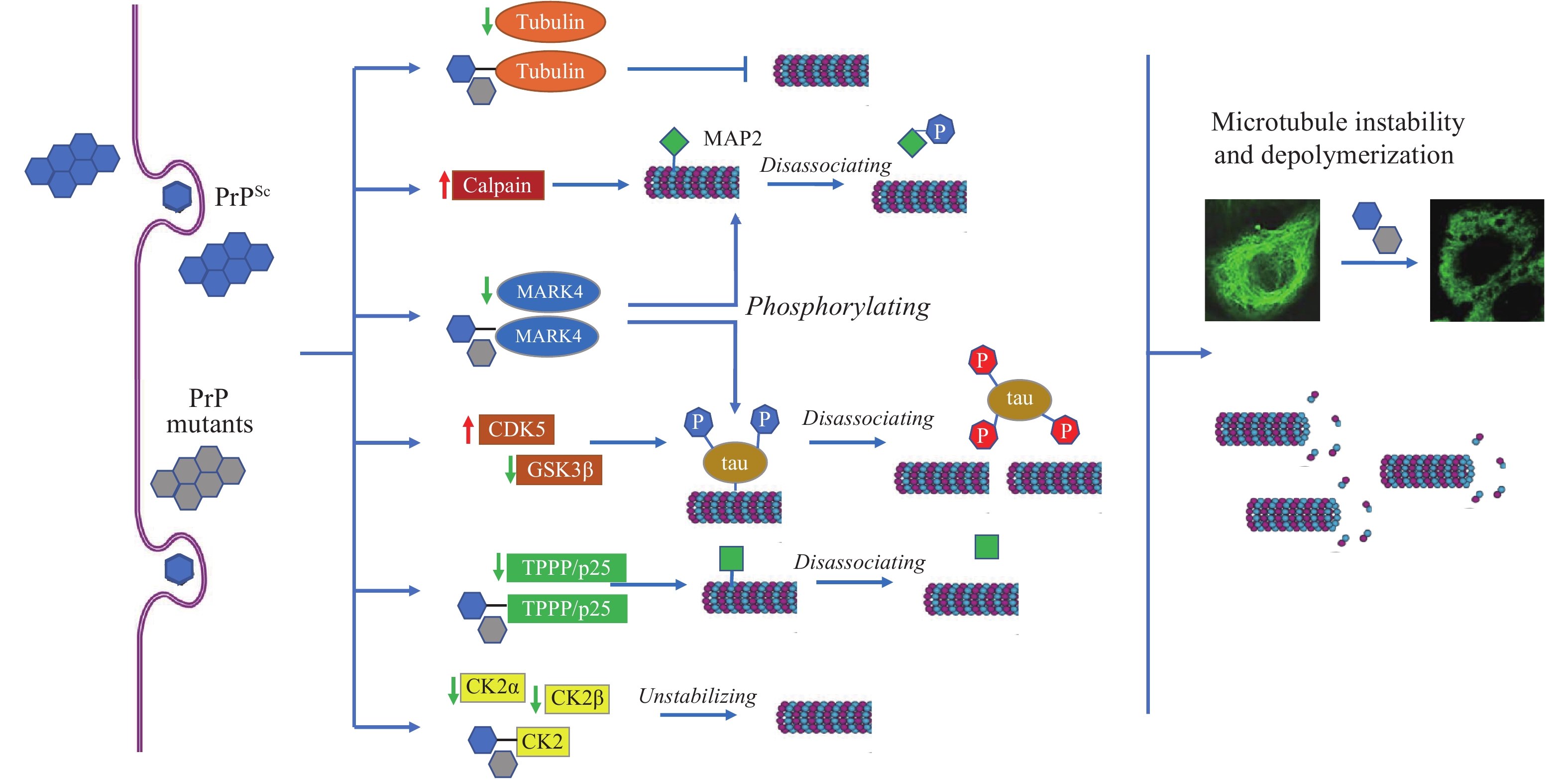
      Figure 1. 

      Schema of prion-induced microtubule instability and depolymerization in brains.

      Note: PrPSc and PrP mutants accumulate in neurons and induce the abnormal changes in tubulin, calpain, and many MAPs, leading to microtubule instability and depolymerization.

      Abbreviation: MAPs=microtubule associated proteins; PrPSc=scrapie-like prion protein; PrP=prion protein, TPPP=Tubulin polymerization promoting protein.

      PrP regulates microtubule dynamics via interacting with microtubule-associated proteins (MAPs). Tubulin polymerization promoting protein/p25 (TPPP/p25) is a brain-specific protein stabilizing cellular microtubular ultrastructure. The brain levels of TPPP of scrapie-infected hamsters were significantly reduced. Distinct molecular interactions between TPPP and PrP were identified. Expression of TPPP in cultured cells significantly antagonized the disruption of microtubule structures and rescued the cytotoxicity caused by the accumulation of Cyto-PrP (14).

      Tau proteins are also important MAPs in the CNS, showing molecular binding capacity with PrP (15). Tau can interact with tubulin and facilitate microtubule formation. In vitro, PrP showed a notable inhibitive effect not only on the interaction of tau with tubulin, but also on the tau-mediated microtubule formation (16). GSS-related mutant P102L and gCJD-related mutants with 2 and 7 extra ORs showed more active binding capacity with tau than wild-type PrP (17). Inhibition of PrP on tau-enhanced formation of microtubule undergoes, at least partially, through blocking the interaction between tau and microtubule.

      Microtubule affinity-regulating kinase 4 (MARK4) is responsible for phosphorylating neuronal MAPs. Significant decreases of brain MARK4 were found in human CJD and scrapie-infected experimental animals, which showed a close correlation with the amounts of PrPSc. Treatment of toxic PrP106-126 peptide onto cultured cells induced a similar reduction of MARK4 and reduction of p-tau at Ser262, indicating that a reduction of MARK4 will result in abnormalities of tau phosphorylation and possibly induce further detachment of microtubules and hinder microtubule transportation (18). We have also identified that MAP2 was markedly decreased in the brains of human prion diseases and scrapie infected rodents. Exposure of the cultured cells to PrP106-126 peptide resulted in a significant reduction of cellular MAP2 and disrupted the microtubule structure. Meanwhile, the level of calpain, which mediates the degradation of a range of cytoskeletal proteins, was significantly increased during prion infection (19).

      Protein kinase CK2 is generally composed of 2 catalytic subunits (CK2α or CK2α’) and 2 regulatory subunits (CK2β) and forms a tetrameric structure. The brain levels of CK2α and CK2β decreased in the scrapie-infected experimental rodent models and human PrDs at the terminal stage (20). Strong molecular interaction was observed between PrP and CK2α subunit (21). Reductions of tubulin and disruptions of microtubule structures induced by abnormal PrP mutants could be completely restored by overexpressing CK2α and CK2β in cells. Strong binding activity of the PrP mutants with CK2 will directly affect the interaction of CK2 with tubulin and other substrates (22).

    • The p21-activated kinases (PAKs) are a group of protein serine/threonine kinases; among them, PAK1 and PAK3 are highly expressed in brain tissues. Remarkable reductions of PAK3 and PAK1 were detected in scrapie-infected brains. The upstream activator Rac/cdc42 and downstream substrate Raf1, particularly the phosphorylated Raf1, were remarkably decreased at terminal stage, strongly highlighting correlation of dysfunction of Rac/cdc42-PAKs-Raf1 signaling with neuron loss during prion infection (23).

      Wnt/β-catenin signaling is critical for synaptic disassembly and neuron loss. In prion infected rodent models, the brain levels of phosphor-β-catenin (Ser33,37 and Thr41) were significantly up-regulated and its target substrate cyclin D1 was down-regulated. Increases of dickkopf-1, the antagonist of Wnt/β-catenin signaling, and decreases of Wnt-3 and phosphor-GSK3β were also found in scrapie-infected brains. Such abnormal changes displayed time-dependent progression along with the incubation period, reflecting a repression of Wnt/β-catenin signaling in neurons (24).

      Polo-like kinases (PLKs) are a family with five major members. PLK3 phosphorylates Cdc25C on Ser216 and inhibits the activity of Cdc25C, while PLK1 phosphorylates Cdc25C on Ser198 and leads to activation and nuclear import, which is vital in synaptic plasticity. The increase of PLK1 and decrease of PLK3 were identified in scrapie-infected brains. Cdc25C and its phosphorylated forms were down-regulated, and Cyclin B1 and PCNA were obviously up-regulated. Aberrant changes of cell cycle regulatory PLKs-Cdc25C-Cyclin B1signaling in the prion-infected neurons lead to the cell cycle arrest at M phase (25).

      Brain-derived neurotrophic factor (BDNF) appears primarily presynaptic effects at central, autonomic, and neuromuscular synapses. The brain level of BDNF was significantly reduced in scrapie-infected hamsters (26). The major components of BDNF signaling were also remarkably decreased, including tropomyosin-related kinase (Trk) receptors, phosphor-TrkB (p-TrkB), growth factor receptor-bound protein 2 (GRB2) and neurotrophin receptor p75 (p75NTR). Remarkable reduction of brain BDNF-TrK-GRB2-p75NTR signaling reflects a severe depletion of neurotrophic factors and a defect of energy metabolism in local microenvironment.

      Nerve growth factor (NGF) is another member of neurotrophin family. A decrease of brain NGF was observed in the prion infected experimental animals and in prion infected cell line (27-28). Its upstream positive regulatory kinases, p-CREB, p-CaMKIV and CaMKK2, were decreased, whereas the negative regulatory one, p-CaMKK2, was increased. The aberrant situations of those kinases in prion infected cell lines could be also partially reversed by removal of prion agent by resveratrol. Prion infection affects the activity of CDK5-CaMKK2-CaMKIV-CREB cascade, which reduces the expression of NGF.

      Glucose transporter 3 (GLUT3) mainly mediates the glucose passing through the membrane of neurons. The brain GLUT3 levels in scrapie-infected rodents and the prion infected cell line were significantly downregulated (29). The level of hypoxia-inducible factor-1 alpha (HIF-1α), which positively regulated the expressions of GLUTs, was also markedly downregulated in the scrapie-infected brains. The glucose uptake activity in the prion infected cell line was markedly decreased. It indicates a severe defect in glucose uptake and metabolism of neurons during prion infection.

    • Abnormal mitochondrial, oxidative and ER related stresses were observed in the prion infected brains and the cultured cells accumulated with PrPSc or PrP mutants, involving numerous brain proteins and pathways (Figure 2). Reliable molecular interaction between PrP and 14-3-3β in vitro and in vivo was illustrated. Wild-type PrP was promoted, but PrP106-126 peptide inhibited the dimerization of 14-3-3β (30). Time-dependent reduction of 14-3-3 was detected in the brains of scrapie-infected mice, accompanied with the increase of its S-nitrosylated form and many mitochondrial apoptotic agents. Challenge of PrP106-126 onto the cultured cells decreased the level of 14-3-3 and induced translocations of cellular Bax to the membrane fractions. Knockdown of 14-3-3 aggravated the mitochondrial apoptosis in PrP106-126 exposed cells (31).

      Figure 2. 

      Schema of prion-induced mitochondrial stress, ER stress, and oxidative stress in brains.

      Note: Accumulations of PrPSc and PrP mutants cause aberrant alterations (either expressing level or activity or translocation) of numerous brain proteins and pathways related to mitochondrial stress, ER stress, and oxidative stress.

      Abbreviation: PrPSc=scrapie-like prion protein; PrP=prion protein; ER=endoplasmic reticulum.

      Increases of ER-stress-related proteins Grp94, XBP1, TRAF2, and CHOP in the cells expressing PrP mutants with extra ORs (PG9 and PG12), while decrease of Bcl-2 and increase of cytochrome C in the cells expressing the mutant removal of ORs (PG0). The mutants with inserted ORs underwent mainly via ER stress and the mutant without OR via mitochondrial-related pathway (32). Accumulation of Cyto-PrP in cytoplasm also induces cell apoptosis, in which mitochondrial apoptotic pathway seems to be essential (33). Dynamin-related protein 1 (Drpl) and optic atrophy protein 1 (Opa1) are two essential elements for mitochondria fission and fusion. Reductions of brain Drp1 and Opa1 were noticed in various scrapie-infected animals, indicating a disturbance of brain mitochondria dynamics (34).

      Swelling mitochondria structures were observed in the prion-infected cell line by transmission electron microscopy. The levels of Pink1 and Parkin, particularly in the mitochondrial fraction, were increased, whereas the levels of mitochondrial membrane proteins TIMM44, TOMM20, and TIMM23 were decreased. The brain Pink1 and Parkin in mice infected with scrapie were also increased at the terminal stage, predominantly located at the areas with more PrPSc deposit. Enhanced Pink-Parkin pathway mediated activated mitophagy during prion infection (35).

      Cells expressing PrP mutants (KDEL and 3AV) produced an 18-kD large C-terminal proteolytic resistant fragment (Ctm-PrP). Those cells were more sensitive to ER stress stimuli, showing ER-mediated apoptosis by CHOP and the caspase-12 apoptosis pathway. Ctm-PrP and ER stress were only observed in the cells expressing PrP mutants in the transmembrane region (G114V and A117V), but not in those expressing the mutants outside the transmembrane region (36). Protein disulfide isomerase (PDI) is an enzymatic chaperone for reconstructing misfolded protein in ER lumen. In the scrapie-infected hamsters, abnormally upregulated brain PDI, Grp78 and Grp58 were detected. Expressions of PrP mutants in the cultured cells induced high levels of PDI and Grp18 and cytotoxicity. PDI and its relevant executors may function as a pleiotropic regulator in the processes of misfolded PrP proteins (37).

      The OR domain was critical for PrP antioxidation. Disruption of ORs induced a high level of ROS and a low level of glutathione peroxidase in cells that were more susceptible to the challenges of oxidative factors (38). Abnormal increases of ROS are repeatedly reported in the brains of human and animal prion diseases. Molecular interaction between PrP and HS-1 associated protein X-1 (HAX-1), a protein associated with cell apoptosis. Co-expression of wild-type PrP and HAX-1 increased the cultured cells to resist the challenge of H2O2 (39).

    • Although the clinical and general neuropathological features of prion disease have been well documented for a hundred years, the molecular abnormalities started to be revealed in the past three decades. Along with the development and progress of molecular biology and omics, the mysterious veil of the pathogenesis of CJD-associated neuropathological changes has been gradually revealed by uncovering the aberrant alterations of dozens of bioactive components and biological pathways. Extensive dysfunctions of numerous biological processes and signals directly lead to slow but irreversible damage on neurons and brain tissues infected with prions, the majority of them occurring much earlier than the appearance of clinical symptoms, even earlier than the appearance of detectable PrPSc. Other neurodegenerative diseases, such as AD and PD, also display similar neuropathological processes. However, besides the unique infectivity of prions, the clinical duration of PrD is much shorter, which possibly reflects a much faster and more severe neuropathological deterioration.

    • All staffs, doctoral and master students in the Department of Prion Disease of National Institute for Viral Disease Control and Prevention, China CDC since 1998 and the colleagues from provincial CDCs and sentinel hospitals in National Surveillance for prion diseases since 2006.

Reference (39)

Citation:

Catalog

    /

    DownLoad:  Full-Size Img  PowerPoint
    Return
    Return