
 Download:


HTML
-
Introduction: Nontuberculous mycobacteria (NTM) and Mycobacterium tuberculosis (MTB) share significant genomic similarity, enabling NTM to induce protective immune responses against MTB infection. This characteristic has led to their increasing application in tuberculosis (TB) vaccine development. This study evaluated the immunological properties of a Mycobacterium intracellulare (Mit) strain to provide scientific evidence for the development of novel TB vaccines.
Methods: Whole-cell proteins were extracted from the Mit strain CHPC 1.5701 and used to establish a mouse immunization model. Key antibody and cytokine parameters were measured to assess immune responses. Additionally, a subcutaneous air pouch model was developed on the dorsal surface of mice to evaluate neutrophil recruitment capacity.
Results: Mice in the experimental group developed high IgG antibody titers (1:921,600±446,351.3) and demonstrated a Th1-type immune response. Post-immunization serum antibodies exhibited cross-reactivity with MTB whole-cell proteins. The subcutaneous air pouch model revealed substantial neutrophil recruitment following antigen challenge.
Conclusions: Mit whole-cell proteins demonstrate potent immunogenicity and cross-reactivity with MTB whole-cell proteins, suggesting potential applications in the immunoprevention and treatment of tuberculosis.
-
Tuberculosis (TB) is a highly hazardous respiratory infectious disease and currently has the highest mortality rate among infectious diseases caused by a single pathogen (1). At present, Bacillus Calmette-Guerin vaccine (BCG) remains the only globally approved TB preventive vaccine. While BCG provides some protection for infants and young children, its efficacy is poor in adolescents and adults, and it cannot prevent latent TB infection (LTBI). Therefore, to achieve the World Health Organization’s (WHO) goal of “ending the tuberculosis epidemic by 2035,” the development of diverse and more effective TB vaccines is urgently needed (2-3).
Nontuberculous mycobacteria (NTM) refer to a large group of Mycobacteria excluding the Mycobacterium tuberculosis (MTB) complex and Mycobacterium leprae. More than 200 NTM species have been identified (4), and some genetically modified NTMs have been utilized in anti-tuberculosis vaccine development (5), including Mycobacterium intracellulare (Mit). One study (6) identified 2,740 homologous genes between Mit and MTB, with 521 of these genes being rich in B cell antigen epitopes. Another study revealed numerous cross-reactive proteins between Mit and MTB (7). Considering that clinical isolates often retain the genetic diversity and biological characteristics inherent in natural infection processes and reflect the characteristics of currently prevalent strains, this study used a clinical Mit isolate to explore its potential for new TB vaccine development.
The clinical Mit strain used in this study was isolated from the sputum of a suspected tuberculosis patient and is preserved at the National Institute for Communicable Disease Control and Prevention, Chinese Center for Disease Control and Prevention. Fresh Mit culture was inactivated in a water bath at 80 °C for 35 min, then centrifuged at 5000 rpm for 10 min. The bacterial cell pellets were washed three times with 0.01M pH 7.2 phosphate-buffered saline (PBS) and resuspended in PBS before being ultrasonically disrupted (250 W, 80 min, 10 s on, 10 s off). Following centrifugation, the supernatant was filtered through a 0.22-μm filter membrane and stored at −80 °C.
Female specific pathogen-free (SPF) BALB/c mice aged 6–7 weeks (Beijing Vital River Laboratory Animal Technology Co., Ltd.) were divided into experimental and control groups, with 5 mice per group.
Mice in the experimental group were immunized subcutaneously three times at two-week intervals with 100 μg of Mit whole-cell proteins per mouse per immunization. The control group received the same volume of sterile PBS. Fourteen days after the final immunization, blood was collected from the orbital sinus, and spleens were aseptically harvested following cervical dislocation.
Antibody levels were determined using conventional ELISA methodology. Whole-cell proteins were diluted in coating buffer (Na₂CO₃-NaHCO₃) to a final concentration of 10 μg/mL. For each well of a 96-well microtiter plate, 100 μL of this solution was added and incubated at 4 °C for 24 hours. The plates were blocked with 5% skim milk solution. Horseradish peroxidase-labeled sheep anti-mouse IgG and IgM antibodies were used as secondary antibodies. Absorbance values were measured at 450 nm using a microplate reader.
Mice were sacrificed and their spleens were aseptically removed. Each spleen was processed by adding 4–5 mL of mouse lymphocyte separation medium and grinding to form a cell suspension. The cells were washed, resuspended in culture medium, and adjusted to a concentration of 1×10⁶ cells/mL. For each well, 100 μL of cell suspension was plated, followed by the addition of either 10 μL of whole-cell protein antigens (200 ng/μL) or PBS as stimulants. The secretion levels of interleukin-4 (IL-4) and interferon gamma (IFN-γ) were subsequently measured using enzyme-linked immunospot (ELISPOT) assay.
Splenic lymphocytes were prepared at a concentration of 1×10⁷ cells/mL, aliquoted, and stimulated with a mixture of stimulants and blockers. For surface markers, fluorescent-labeled antibodies against cluster of differentiation 4 (CD4), CD8, CD62L, and CD44 were used. For cytoplasmic antigens, fluorescent-labeled antibodies against interleukin-2 (IL-2), IL-4, and IFN-γ were employed. Analysis was performed using flow cytometry.
Lymphocytes (1×10⁵ cells) were seeded in 96-well cell plates and incubated with 5 μg of whole-cell proteins for 24 hours. The cell culture was centrifuged at 300 g for 10 minutes at 4°C, and the supernatant was collected. The remaining culture was further centrifuged at 3,000 g for 10 min at 4 °C to collect additional supernatant. These collected supernatants were analyzed for cytokine levels using the Luminex 200 system (Luminex Corporation, Austin, TX, USA).
A subcutaneous air pouch model was established by injecting 4 mL of sterile air into the dorsal region of mice, followed by an additional 3 mL injection at the same site three days later. On day 6, 1 mL of 0.5% carboxymethyl cellulose (CMC) solution, with or without the whole-cell proteins, was injected into the air pouch. Mice were sacrificed 24 hours after injection. Flow cytometry was used to characterize immune cell populations, with CD11b+Ly6GhiF4/80− cells identified as neutrophils and CD11b+F4/80hiLy6G− cells identified as macrophages.
Mouse sera with the highest antibody titers were pooled and used for cross-immunization testing with an MTB whole-proteome chip (BC Biotechnology Co., Ltd) (8). The detailed test procedures have been described in a previous publication (7). Functional clustering analysis of the reactive proteins was performed according to Gene Ontology (GO) and Kyoto Encyclopedia of Genes and Genomes (KEGG).
The results demonstrated that mice in the experimental group developed high antibody titers, with IgG, IgG1, and IgG2a reaching 1:921,600±446,351.3, 1∶32,000±11,085.1, and 1∶67,200±37,727.4, respectively (
Supplementary Figure S1 ).The results demonstrated that the production level of Th1-type cytokine IFN-γ was significantly higher than that of Th2-type cytokine IL-4 (P<0.01), suggesting that the immune response generated by splenic lymphocytes from immunized mice following stimulation with whole-cell protein antigens was predominantly Th1-biased (Figure 1).
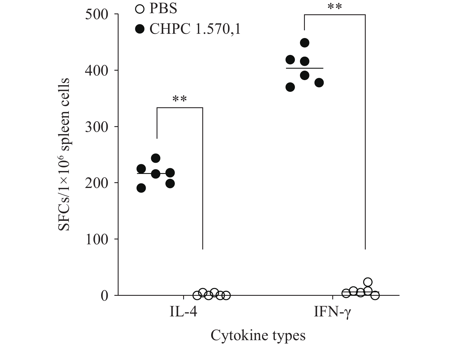 Figure 1.
The number of spots of IL-4 and IFN-γ secreted by splenic lymphocytes of mice stimulated with the whole-cell protein antigens of CHPC 1.5701.
Abbreviation: CHPC=the Center for Human Pathogen Collection; PBS=phosphate-buffered saline; IFN-γ= Interferon-γ; IL-4=Interleukin-4; SFCs=spot-forming cells.
** P<0.01.
The results demonstrated that the numbers of IL-2+CD4+, TNF-α+CD4+, and TNF-α+CD8+ T lymphocytes in the experimental group were significantly higher than those in the control group (P<0.05) (Figure 2A). Similarly, multifunctional CD4+ T lymphocytes expressing IFN-γ+TNF-α+ and TNF-α+IL-2+, as well as CD8+ T lymphocytes expressing IFN-γ+TNF-α+IL-2+ and IFN-γ+TNF-α+, were also significantly elevated compared to the control group (Figure 2B and Figure 2C).
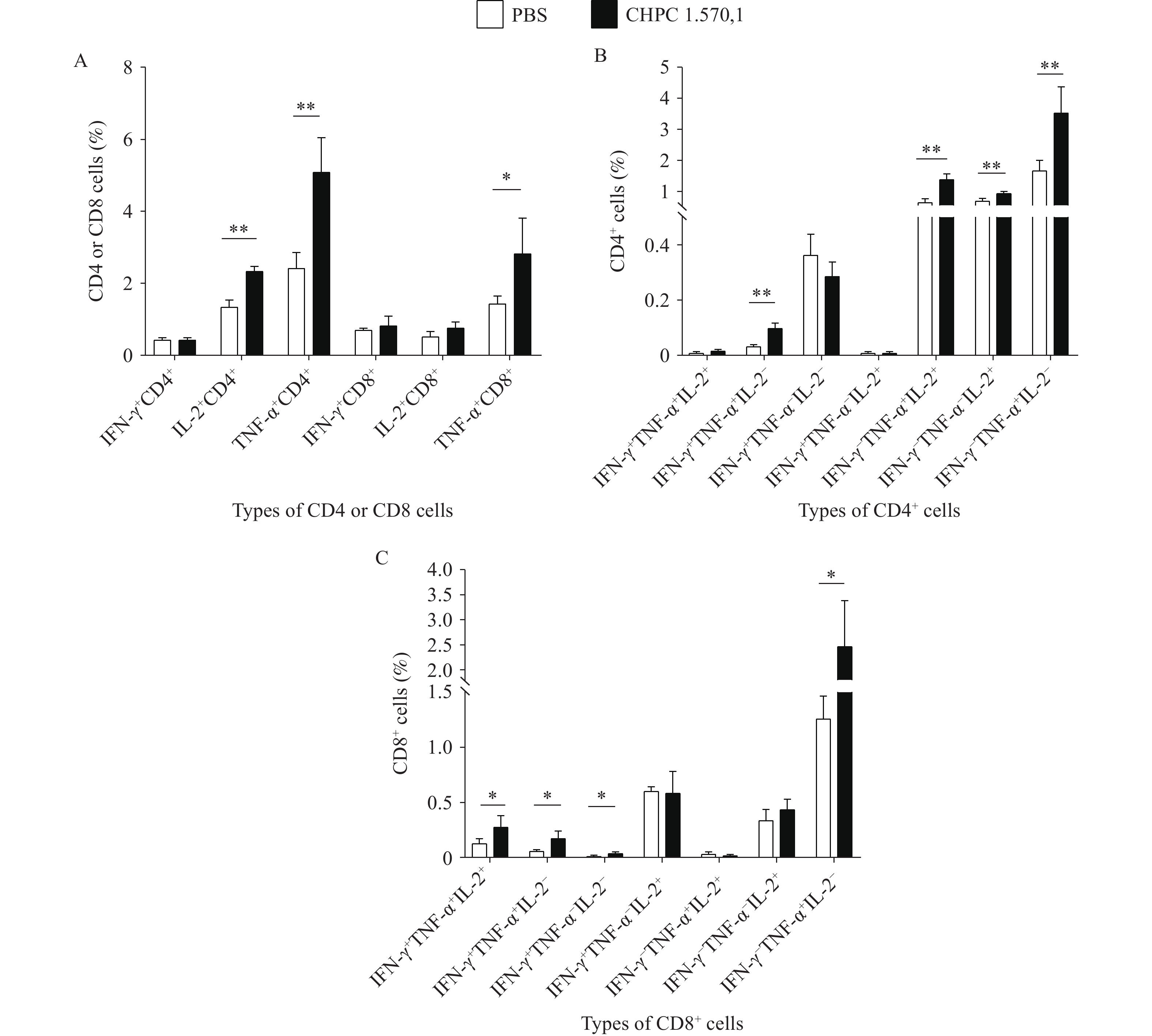 Figure 2.
The results of the level of cellular immune response. (A) The numbers of IL-2+CD4+, TNF-α+CD4+, and TNF-α+CD8+ T lymphocytes; (B) The level of IFN-γ, TNF-α, and IL-2 secreted by multifunctional CD4+ T lymphocytes in mice; (C) The level of IFN-γ, TNF-α, and IL-2 secreted by multifunctional CD8+ T lymphocytes in the mice.
Abbreviation: CHPC=the Center for Human Pathogen Collection; PBS=phosphate-buffered saline; IFN-γ=Interferon-γ; TNF-α=Tumor necrosis factor-α; IL=Interleukin; CD=Cluster of differentiation.
* P<0.05.
** P<0.01.
Compared with the control group, the levels of CD4+TCM, CD8+TCM, CD4+TEM, and CD8+TEM in the experimental group increased to varying degrees. Among these, IL-2+CD4+TCM, CD4+TEM, IFN-γ+CD8+TEM, and TNF-α+CD8+TEM increased significantly, indicating that immunization with the whole-cell protein antigens of Mit can enhance the proliferation of central memory T cells (TCM) and effector memory T cells (TEM) in mice (Figure 3).
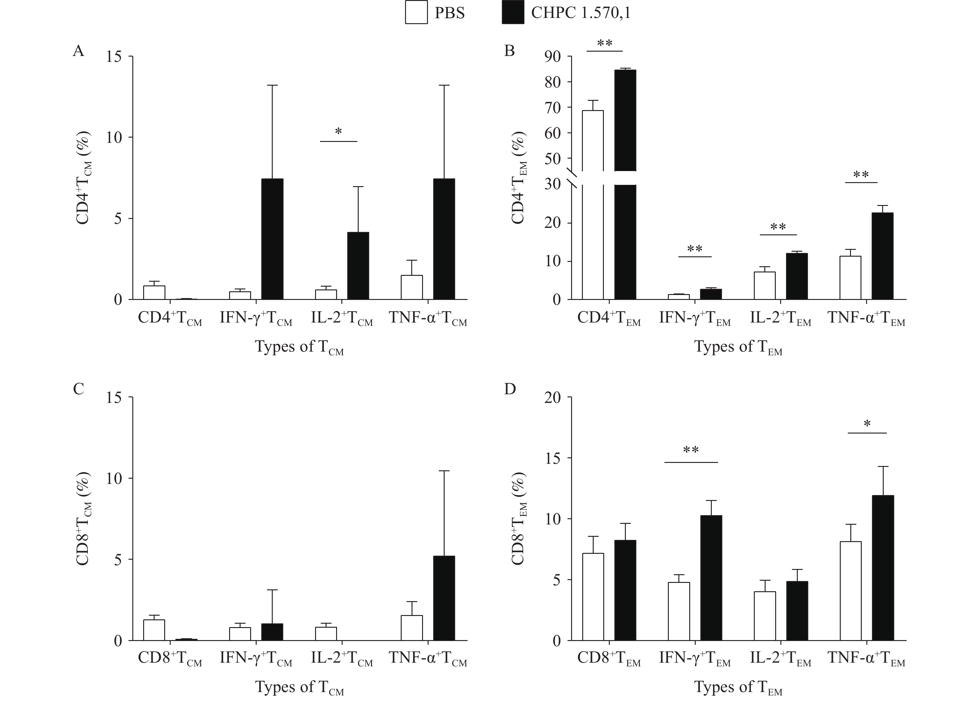 Figure 3.
The results of the number of TCM and TEM cells in splenic lymphocytes of mice after immunization with the whole-cell proteins.(A) Changes in CD4+TCM cells. (B) Changes in CD4+TEM cells. (C) Changes in CD8+TCM cells. (D) Changes in CD8+TEM cells.
Abbreviation: CHPC=the Center for Human Pathogen Collection; PBS=phosphate-buffered saline; IFN-γ=Interferon-γ; TNF-α=Tumor necrosis factor-α; IL-2=Interleukin-2; CD=Cluster of differentiation; TCM=central memory T cells; TEM=effector memory T cells.
* P<0.05.
** P<0.01.
The detection results of Th1, Th2, and Th17-type cytokines revealed that after stimulation with MTB whole-cell protein antigens, the levels of IFN-γ, IL-2, IL-4, IL-6, IL-10, IL-17A, and GM-CSF in the experimental group increased significantly compared to controls. These findings indicate that immunization with Mycobacterium intracellulare whole-cell protein antigens significantly enhances the secretion of cytokines that inhibit MTB proliferation (Figure 4).
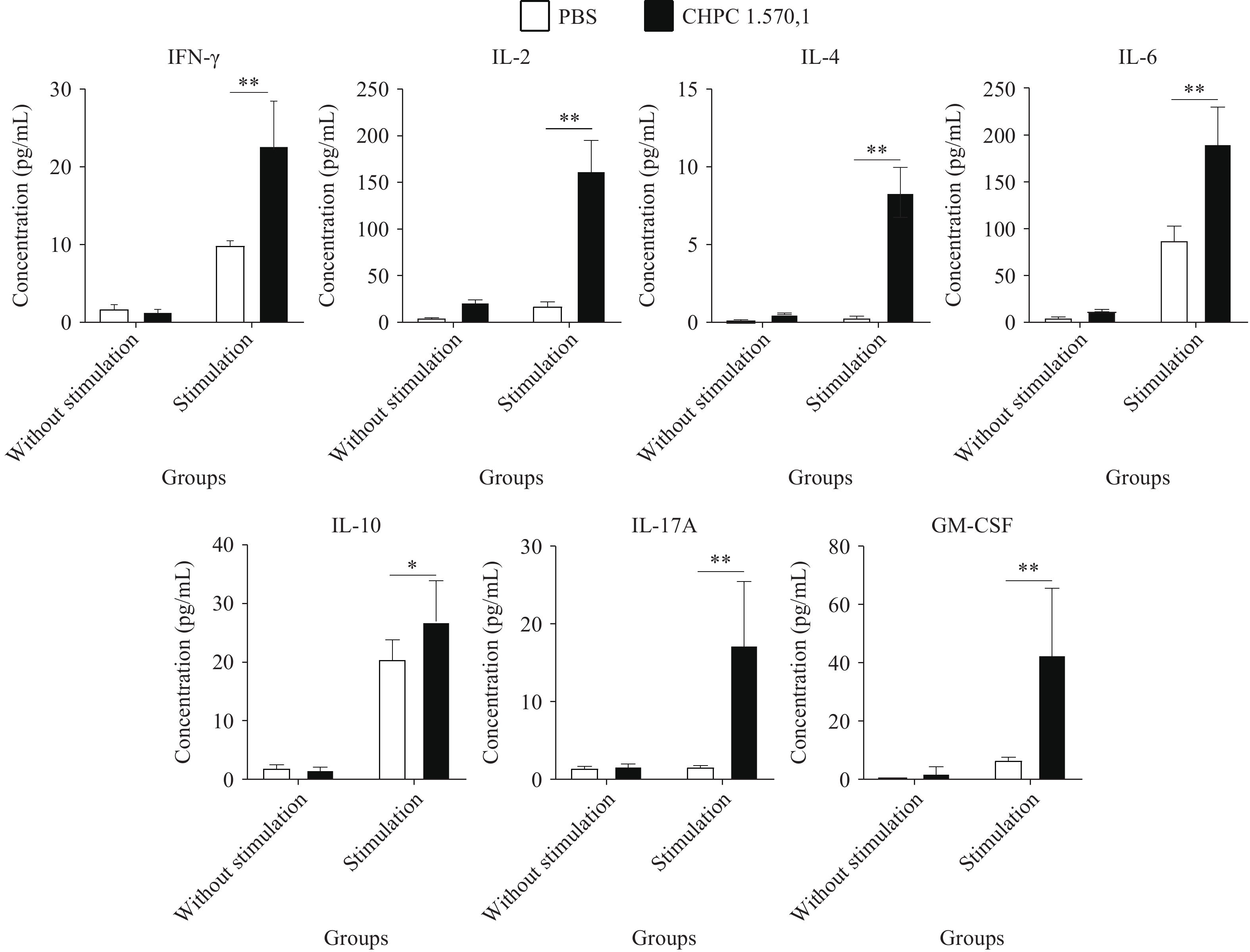 Figure 4.
The results of the levels of cytokines secreted in splenic lymphocytes of the mice stimulated by MTB whole-cell protein antigens.
Abbreviation: CHPC=the Center for Human Pathogen Collection; PBS=phosphate-buffered saline; IFN-γ=Interferon-γ; IL=Interleukin; GM-CSF=Granulocyte Macrophage Colony-Stimulating Factor.
* P<0.05.
** P<0.01.
Flow cytometry analysis revealed that 24 hours after injection of Mit whole-cell protein antigens, the antigen-injected group recruited significantly more CD11b+ cells (primarily inflammatory cells including neutrophils, monocytes, and macrophages) compared to the control group injected with CMC alone (
Supplementary Figure S2A ). Additionally, a substantial number of neutrophils were recruited to the injection site (Supplementary Figure S2B ).The Results of Cross-Immunization Test between Immune Serum of the Mice Immunized with whole-cell protein antigens and MTB Whole-Proteome Chip
The serum from the experimental group of mice was tested against the MTB whole-proteome chip containing 4,262 recombinant proteins to identify reactive proteins and indirectly evaluate the post-immunization antibody profile. Results revealed that serum from mice immunized with Mit whole-cell protein antigens recognized a total of 630 MTB proteins.
KEGG and GO functional analyses were performed on the positive proteins. Results showed that these proteins were involved in various biological processes including metabolic pathways, nucleic acid metabolism, protein export, biosynthesis of amino acids, biosynthesis of secondary metabolites, cytoplasmic membrane functions, ATP binding, and protein folding.
-
In this study, we constructed a mouse immunization model to evaluate the immunological properties of Mit, including humoral immunity, cellular immunity, and anti-MTB infection responses. The whole-cell protein antigens from the Mit strain CHPC 1.5701 elicited a stronger humoral immune response in mice compared to previous studies (6-7), enhanced cellular immune responses, and demonstrated significant neutrophil recruitment capability. These findings provide valuable insights for tuberculosis vaccine development.
In recent years, NTM have emerged as an important research focus for TB vaccine development. Studies have shown that the rBCG30 recombinant BCG vaccine enhances immunoprotective effects in animal models (9). Additionally, the subunit vaccine ID93/GLA-SE, which targets common antigens shared between NTM and MTB, has progressed to clinical trials for safety and efficacy evaluation (10). Our findings with this clinical isolate demonstrate favorable cross-immune responses between NTM and MTB. Furthermore, the whole-cell proteins of Mit CHPC 1.5701 exhibited significant neutrophil recruitment within 24 hours after injection in mice, suggesting potential to strengthen the first line of defense against MTB infections.
However, our study has limitations. During animal model establishment, we only included control and experimental groups injected with whole-cell proteins, without examining the immune response effects of Mit CHPC 1.5701 combined with an adjuvant. Additionally, our humoral immunity assessment was limited to IgG antibody detection and did not include other antibody levels. Future research should address these limitations to obtain more comprehensive information.
In conclusion, our findings on this clinical isolate not only expand our understanding of the relationship between Mit and MTB but also identify a potential strain for tuberculosis vaccine development.
-
Approved by the Ethics Committee of the National Institute for Communicable Disease Control and Prevention, Chinese Center for Disease Control and Prevention (No. 2022-025).
| Citation: |

|