
 Download:


-
Introduction: Echinococcosis is a zoonotic parasitic disease caused by the larval stage of Echinococcus, prevalent in northwestern China. It poses a serious threat to human health and causes significant economic losses in the livestock industry. This study aims to investigate the infection and development of Echinococcus in livestock in northwestern China, providing scientific basis for precise prevention and control of echinococcosis.
Methods: This study utilized a combination of slaughterhouse and household investigations. Liver and lungs from slaughtered livestock in Sichuan Province, Qinghai Province, Xizang Autonomous Region, and Xinjiang Uygur Autonomous Region were examined through visual inspection and palpation, and Echinococcus cysts were collected. The cyst fertility was analyzed via microscopic examination. Metacestode DNA was extracted for PCR amplification of the mitochondrial Cox1 gene. Sequence alignment with the GenBank database was conducted to identify the genotypes of Echinococcus. Phylogenetic tree was constructed using MEGA 7.0 software. Haplotypes were analyzed using DnaSP 6, and a haplotype network was constructed using PopART 1.7. Data analysis was performed using SAS 9.4, and P<0.05 indicates statistical significance.
Results: Between October and December 2023, 400 yaks and 808 sheep were surveyed in Qinghai, Xizang, and Xinjiang. The infection rate of Echinococcus in yaks was 16.5%, significantly higher than that in sheep (9.41%, χ2=12.9802, P<0.001). The fertility rate of Echinococcus cysts in sheep was 71.79%, significantly higher than that in yaks (15.57%, χ2=64.1670, P<0.0001). The cysts were mainly parasitizes in the liver of sheep (82.89%) and the lungs of yaks (63.89%). A total of 169 Cox1 sequences were successfully amplified, of which 98.82% (167/169) corresponded to E. granulosus sensu stricto (s.s.) G1/G3, while one sequence was identified as E. canadensis G6, and one from E. multilocularis. A total of 48 haplotypes were detected, with H3 being the predominant haplotype.
Conclusions: In the four survey provincial-level administrative divisions (PLADs) of China, the infection rate in yaks (16.5%) was significantly higher than in sheep (9.41%). Echinococcus was found preferably infect sheep liver and yak lungs, with a higher cyst fertility rate in sheep compared to yaks. Livestock infections are mainly caused by E. granulosus G1/G3, and this study, for the first time, identified E. multilocularis infection in yaks from Xizang. The findings provide a crucial foundation for further research into the molecular epidemiology, genetic evolution, and the development of precise prevention and control strategies for Echinococcus in the regions.
-
Echinococcosis is a serious and potentially fatal parasitic zoonosis with worldwide distribution, caused by the larval stage of cestodes in the genus Echinococcus. Cystic echinococcosis (CE) caused by Echinococcus granulosus sensu lato (s.l.) and alveolar echinococcosis (AE) caused by E. multilocularis are the two main forms of the disease and represent major public health problems in northwestern China. E. granulosus s.l. is a species complex comprising E. granulosus sensu stricto (s.s.) (G1/G3), E. equinus (G4), E. ortleppi (G5), E. canadensis (G6, G7, G8, and G10), and E. felidis. Among these, G1/G3 and G6/G7 are commonly found in human infections, while the other genotypes are either rare or absent in human cases (1). E. granulosus s.l. can infect wild mammals and domestic livestock, while humans may become infected through accidental ingestion of infective eggs (2). The infection leads to the development of Echinococcus cysts primarily in the host’s liver or lungs (3-4). The larval metacestodes can develop into fertile cysts containing infectious protoscoleces, thereby promoting the cycle and transmission of Echinococcus between intermediate and definitive hosts. However, field investigations frequently reveal infertile and calcified cysts that cannot continue the parasite life cycle, thus reducing transmission risk in local areas. In China, developmental differences in cysts among intermediate hosts remain poorly understood, which impacts the risk assessment of Echinococcus transmission. Understanding the genotype distribution and cyst development patterns in different hosts is crucial for implementing effective prevention and control strategies against echinococcosis.
This study was conducted in four provincial-level administrative divisions (PLADs) in northwestern China: Xinjiang Uygur Autonomous Region, Sichuan Province, Qinghai Province, and Xizang Autonomous Region, with average altitudes ranging from 1,500 to 4,700 meters. From October to December 2023, livestock infection investigations were conducted at slaughterhouses in Shiquan County, Sichuan; Yushu County, Qinghai; and Hejing County, Xinjiang, as well as in herders’ homes in Mozhugongka County and Dangxiong County, Xizang. Livestock cysts were examined and collected through autopsy. Cyst fertility was evaluated microscopically: the presence of protoscoleces indicated a fertile cyst, while their absence indicated an infertile cyst (Figure 1A). DNA was extracted from cysts using the DNeasy Blood and Tissue Kit (Qiagen, Hilden, Germany) following the manufacturer’s instructions. The primers forward: TTGAATTTGCCACGTTTGAATGC and reverse: GAACCTAACGACATAACATAATGA were used to amplify an 874 bp fragment of the cytochrome c oxidase subunit 1 (Cox1) gene. Polymerase chain reaction (PCR) reaction conditions followed the protocol described by Minoru Nakao et al (5). CE prevalence, cyst fertility, and organ preference were compared using χ2 test or Fisher’s exact test, with differences considered statistically significant at P<0.05. All statistical analyses were performed with SAS (version 9.4; SAS Institute; Cary, North Carolina, United States). All amplicons were sequenced and compared to sequences in GenBank (https://blast.ncbi.nlm.nih.gov/Blast.cgi). A phylogenetic tree based on Cox1 sequences was constructed using MEGA (version 7.0; Center for Evolutionary Medicine at Temple University; Philadelphia, Pennsylvania, United States) and visualized with iTOL (https://itol.embl.de/). Haplotype diversity was analyzed with DnaSP (version 6) (6). The haplotype network and statistical calculations were performed using the Median-Joining network method in PopART software (version 1.7) (7).
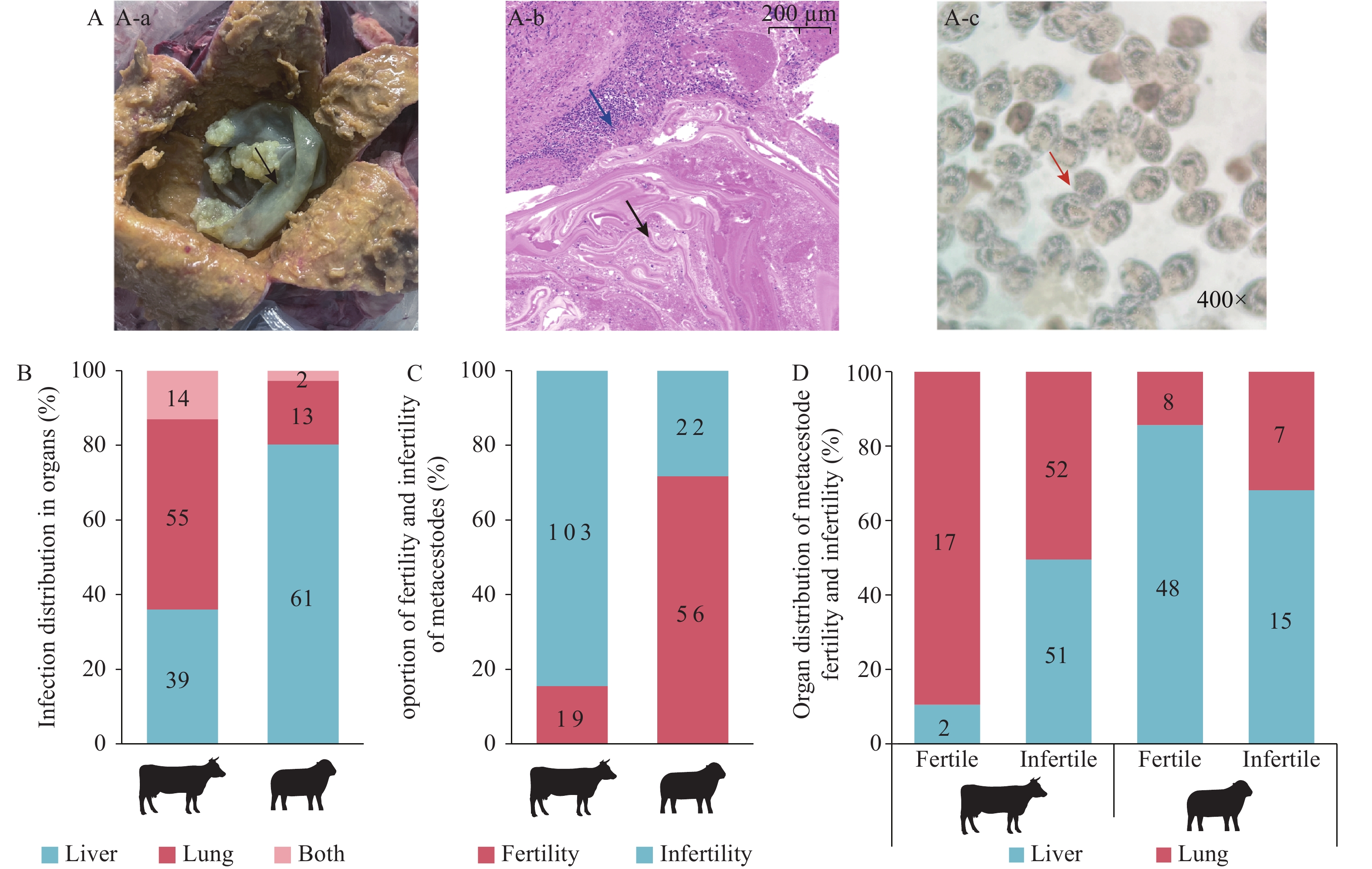 Figure 1.
Metacestode fertility analysis. (A) Autopsy and microscopy examination. A-a: Cyst after fluid extraction; A-b: Histological analysis of cyst stained with haematoxylin-eosin (HE); A-c: Protoscoleces under the microscope. (B) Organ preference of Echinococcus in yaks and sheep; (C) Comparison of metacestode fertility rates between yaks and sheep. (D) Comparison of metacestode fertility rates in different organs of yaks and sheep.
Note: black arrow - endocyst; blue arrow - inflammatory reaction; red arrow - protoscoleces.The present study investigated the prevalence of echinococcosis in livestock across three PLADs. A total of 400 yaks (8–14 years old) and 808 sheep (2–5 years old) were examined. We identified 158 cysts across three PLADs, including 32 cysts in yaks from Qinghai, 48 cysts in yaks from Xizang, 20 cysts in sheep from Xizang, and 58 cysts in sheep from Xinjiang. The infection rate in yaks (16.5%, 66/400) was significantly higher than in sheep (9.41%, 76/808, χ2=12.9802, P<0.001). Notably, the prevalence of cysts in Xizang yaks (43.59%, 34/78) was significantly higher than in Qinghai yaks (9.94%, 32/322, χ2=51.6105, P<0.0001). Similarly, the prevalence in sheep from Xinjiang (17.06%, 58/340) was significantly higher than in Xizang (3.85%, 18/468, χ2=40.3453, P<0.0001). Additionally, 42 cysts from yaks in Sichuan and 5 cysts from cattle in Xinjiang were included in the genotype and fertility analyses.
Cox1 genes were successfully amplified in 169 samples, with an overall success rate of 82.44% (169/205), comprising 78.69% (96/122) from yak cysts and 89.74% (70/78) from sheep cysts. Genotyping revealed that 167 isolates (98.82%) were identified as E. granulosus s.s. (G1/G3) from Qinghai (n=23), Xizang (n=52), Xinjiang (n=59), and Sichuan (n=33). One isolate was identified as E. canadensis (G6) from sheep, and one as E. multilocularis from yak. Three species/genotypes (G1/G3, G6, and E. multilocularis) were detected in Xizang, whereas only G1/G3 was found in the other three PLADs (Table 1). We identified 46 distinct haplotypes from G1/G3 samples. To explore relationships between different haplotypes, a Median-Joining Network was constructed (Figure 2). The network displayed a star-like structure with one predominant haplotype (Haplotype 3, H3), which accounted for 48.50% (81/167) of the total population and was distributed across all four PLADs. Among the haplotypes, 40 were exclusive to a single PLADs, while 5 haplotypes were prevalent in two geographically adjacent PLADs. E. granulosus s.s. shared haplotypes between yaks and sheep. Specifically, H3 was shared across all four PLADs, while haplotypes H9, H28, H31, H32, H34, and H37 were shared in Xizang.
PLAD Host No. of
examinationInfection rate, % No. of
cystsFertile cyst
rate, %Genotype No. Of
isolateQinghai Yak 322 9.94 (32/322) 32 3.13 (1/32) G1/G3 23 Xizang Yak 78 43.59 (34*/78) 48 29.17 (14/48) G1/G3 39 E. multilocularis 1 Sheep 468 3.85 (18*/468) 20 35.00 (7/20) G1/G3 13 G6 1 Xinjiang Sheep 340 17.06 (58/340) 58 84.48 (49/58) G1/G3 56 Cattle 5 0 G1/G3 3 Sichuan Yak 42 9.52 (4/42) G1/G3 33 Total Yak 400 16.5 (66/400) 122 15.57 (19/122) G1/G3 95 E. multilocularis 1 Sheep 808 9.41 (76/808) 78 71.79 (56/78) G1/G3 69 G6 1 Cattle 5 0 G1/G3 3 Note: The four PLADs include Qinghai, Xizang, Xinjiang, and Sichuan. Infection rate (%) = Number of infected livestock ÷ Number of inspected livestock × 100% ; Fertile cyst rate (%) = Number of fertilecyst ÷ Number of cyst ×100%; No. of cysts was calculated based on the number of infected organs, with each infected organ being counted as one cyst.
Abbreviation: PLADs=provincial-level administrative divisions.
* indicates Echinococcus infection in multiple organs, including the liver and lung.Table 1. Prevalence, genotype diversity and cyst fertility of Echinococcus isolates from 4 PLADs in China.
 Figure 2.
Haplotype network and phylogenetic analysis of the 735 bp Cox1 gene of Echinococcus. (A) Haplotype network: Circle sizes represent the frequency of each haplotype. (B) Phylogenetic analysis: Different haplotypes belonging to the same species/genotype are colored accordingly.
Note: For (A), The number of mutations separating haplotypes is indicated by dash marks. Colors indicate the geographic origin. H: haplotype. Circle in blue corresponds to Sichuan (Number of haplotypes, n=16), circle in green corresponds to Qinghai (n=7), circle in purple corresponds to Xizang (n=18), and circle in yellow corresponds to Xinjiang (n=13). For (B), Intermediate hosts for a specific haplotype are indicated by black host silhouette.In this study, the distribution of Echinococcus cysts in the parasitized organs of yaks and sheep showed statistically significant differences (χ2=35.2831, P<0.0001). A total of 122 cysts were found in 108 yaks, including 69 cysts (63.89%) in the lung and 53 cysts (49.07%) in the liver. Multi-organ (both liver and lung) infections were found in 14 yaks. There was a significant difference in the distribution of Echinococcus cysts in yak organs (χ2=4.8218, P<0.05). Meanwhile, a total of 78 cysts were identified from 76 sheep, including 15 cysts (19.74%) in the lung and 63 cysts (82.89%) in the liver. Multi-organ infections were found in 2 sheep. The distribution of Echinococcus cysts in sheep organs was significantly different (χ2=60.6736, P<0.0001). These results indicate that Echinococcus shows a significant organ preference for the lungs in yaks and for the liver in sheep. Additionally, the fertility rates of cysts formed in the parasitized organs were compared. The fertility rate in sheep (71.79%, 56/78) was higher than that in yaks (15.57%, 19/122), with a statistically significant difference (χ2=64.1670, P<0.0001). In sheep, the fertility rate was 76.19% (48/63) in the liver and 53.33% (8/15) in the lungs, with no statistically significant difference. In yaks, the fertility rate was 3.77% (2/53) in the liver and 24.64% (17/69) in the lungs, indicating that the cyst fertility differed significantly (χ2=9.9241, P<0.05) in the distribution of organs in yaks, with Echinococcus more likely to develop infertile cysts in the yak liver (Figure 1B–D).
-
Differences in susceptibility to Echinococcus infection among various hosts can significantly influence parasite growth and development. Understanding these host-parasite interactions is crucial for comprehending echinococcosis epidemiology and implementing effective control measures. Therefore, investigating Echinococcus infection and development in livestock across four PLADs provides essential information for designing accurate, targeted prevention and control strategies.
In this study, CE prevalence was 16.5% in yaks and 9.41% in sheep. The prevalence in Xizang yaks (43.59%) was significantly higher than the national average of 5.8% reported from 2016 to 2021 (4), highlighting the need for continued and enhanced control measures in this region. Conversely, the prevalence in sheep was lower than the post-2011 prevalence (13.86%) reported in a meta-analysis (3). This reduction in sheep prevalence can largely be attributed to the earlier implementation of intervention measures and increased efficacy of vaccines against echinococcosis in sheep (8). Furthermore, the yaks (8–14 years old) examined in this study were generally older than the sheep (2–5 years old), likely contributing to their extended exposure to contaminated environments and increased susceptibility to infection. Our survey identified three Echinococcus genotypes (G1/G3, G6, and E. multilocularis), with G1/G3 being the most prevalent across all four PLADs, consistent with previous findings in China (9-10). A case of E. multilocularis infection was found in a yak from Xizang. Although E. multilocularis infections in yaks and sheep in the Qinghai-Tibet Plateau have been previously reported (11-12), this is the first report of E. multilocularis infection in a Xizang yak, confirmed by PCR sequencing. Therefore, its impact on livestock should be considered in AE prevention and control efforts. Haplotype analysis identified H3 as the dominant haplotype, consistent with earlier studies (10). Additionally, our results show that sheep and yaks share the H3 haplotype, confirming transmission of this haplotype among livestock across the four PLADs.
Organ or tissue preferences are essential for the establishment, survival, and pathogenesis of many parasites (13). Previous studies (14) have demonstrated that E. granulosus G1/G3 preferentially parasitizes the liver of sheep. In our study, E. granulosus showed a clear predilection for sheep liver (84.06%) and yak lungs (67.06%). Additionally, the proportion of fertile cysts was significantly higher in sheep compared to yaks (Figure 1C). These findings suggest that the liver preference and higher cyst fertility of E. granulosus in sheep contribute more substantially to CE transmission than in yaks. However, the role of yaks should not be overlooked, particularly considering their high infection prevalence in Xizang. Despite these observations, the mechanisms underlying organ tropism and cyst fertility of E. granulosus remain incompletely understood. It is still unclear whether these patterns are driven by parasite factors, host factors, or a combination of both. Further investigation into the evolution, parasitism, and pathogenicity of E. granulosus may provide insights into this issue. Moreover, reliance on the mitochondrial Cox1 gene alone is insufficient to fully understand E. granulosus’s adaptation to endemic environments and its development in various hosts. More comprehensive analysis involving whole genome sequencing is needed. Currently, the control strategy for echinococcosis in the four PLADs of China primarily follows an integrated approach, emphasizing dog deworming, livestock vaccination, and centralized slaughtering. However, this survey revealed significant differences in infection rates, Echinococcus species distribution, and cyst fertility in yaks and sheep across the four PLADs. Therefore, we recommend developing region-specific control strategies based on local epidemiological data. In particular, molecular epidemiological monitoring should be implemented to investigate variations in Echinococcus populations across different regions, providing data support for the precise control of echinococcosis. This study has certain limitations: the sampling locations may not fully represent the overall situation in the four PLADs, and the sample size was constrained by the availability of livestock for autopsy.
In summary, this study investigated the prevalence of Echinococcus in livestock across four PLADs. The results showed that E. granulosus infection in Xizang yaks remains higher than the national average. Additionally, E. multilocularis infection was found in Xizang yaks for the first time. E. granulosus G1/G3 is the most prevalent genotype across the four PLADs, with H3 as the dominant haplotype shared by sheep and yaks in these areas. Furthermore, E. granulosus exhibits a clear organ tropism for the liver in sheep and the lungs in yaks, with higher cyst fertility in sheep. This study provides crucial evidence and a foundation for developing more accurate echinococcosis prevention strategies.
-
The staff from the Disease Control & Prevention Centers of Yushu Prefecture, Qinghai Province; Tibetan Autonomous Prefecture of Ganzi, Sichuan Province; and Hejing County, Bayingolin Mongolian Autonomous Prefecture, Xinjiang Uygur Autonomous Region.
HTML
| Citation: |

|